Vagus Nerve Stimulation for Parkinson's Disease: Emerging Evidence
Introduction: Neuroinflammation and Parkinson's Disease
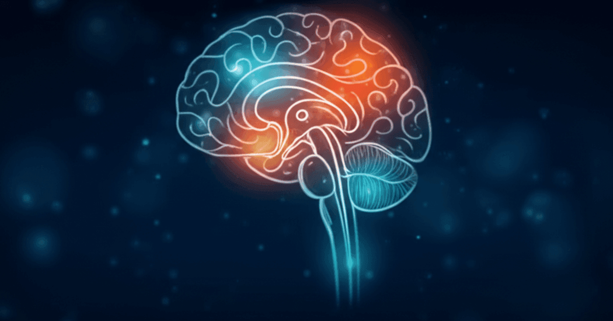
Parkinson's disease (PD) is the second most common neurodegenerative disorder worldwide, affecting approximately 10 million people. It is characterised by the progressive loss of dopaminergic neurons in the substantia nigra pars compacta, leading to the hallmark motor symptoms of tremor, rigidity, bradykinesia, and postural instability. Beyond motor impairment, PD is increasingly recognised as a multisystem disorder involving cognitive decline, autonomic dysfunction, mood disturbances, and gastrointestinal symptoms.
While the precise aetiology of PD remains incompletely understood, a growing body of evidence points to neuroinflammation as a central driver of disease progression. Activated microglia, elevated pro-inflammatory cytokines (including TNF-alpha, IL-1beta, and IL-6), and infiltrating peripheral immune cells have been consistently identified in the substantia nigra of PD patients at post-mortem examination (Tansey & Goldberg, 2010). This inflammatory milieu is thought to create a self-perpetuating cycle of neuronal damage and further inflammation, accelerating the neurodegenerative process.
It is this inflammatory component that has drawn researchers to consider vagus nerve stimulation (VNS) as a potential therapeutic approach for PD. The vagus nerve's well-established role in modulating systemic and central inflammation — through the cholinergic anti-inflammatory pathway — offers a rationale for intervening in a disease process that current treatments can only partially address.
Why VNS? The Therapeutic Rationale
The Cholinergic Anti-Inflammatory Pathway
The primary mechanism linking VNS to potential benefit in PD is the cholinergic anti-inflammatory pathway (CAP). First described by Tracey (2002), this neuroimmune circuit enables the vagus nerve to regulate peripheral and central inflammation. Stimulation of the vagus nerve activates this pathway, leading to suppression of pro-inflammatory cytokine production — particularly TNF-alpha, which has been specifically implicated in dopaminergic neuronal death in PD.
The relevance of this pathway to PD is supported by several converging lines of evidence. First, PD is associated with reduced vagal tone, as measured by heart rate variability, suggesting that the body's endogenous anti-inflammatory brake may be weakened in PD patients (Alonso et al., 2015). Second, the inflammatory markers elevated in PD — TNF-alpha, IL-6, and IL-1beta — are precisely those suppressed by vagal activation. Third, epidemiological studies have found that patients who have undergone vagotomy (surgical severing of the vagus nerve) may have an altered risk profile for PD, though the direction and interpretation of this association remain debated (Svensson et al., 2015; Liu et al., 2017).
Vagal-Dopaminergic Connections
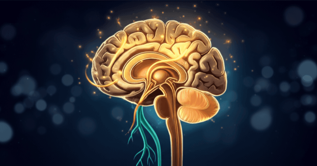
Beyond the anti-inflammatory pathway, there are direct anatomical connections between vagal afferent projections and dopaminergic circuits. The vagus nerve projects to the nucleus tractus solitarius (NTS), which in turn connects to the locus coeruleus and ventral tegmental area (VTA) — a major source of dopaminergic neurons. Preclinical evidence suggests that VNS can modulate dopamine release and metabolism in brain regions affected by PD (Manta et al., 2009), raising the possibility that VNS could influence the very neurotransmitter system that is depleted in the disease.
The Gut-Brain Axis and the Braak Hypothesis
One of the most compelling theoretical links between VNS and PD comes from the Braak hypothesis. Braak et al. (2003) proposed that PD pathology — specifically the accumulation of alpha-synuclein aggregates (Lewy bodies) — may originate in the enteric nervous system and propagate to the brain via the vagus nerve. This "gut-first" hypothesis is supported by observations that gastrointestinal symptoms, including constipation, often precede motor symptoms by years or even decades.
If the vagus nerve serves as a conduit for disease spread, the question arises: could modulating vagal activity through VNS influence this process? While speculative, some researchers have proposed that VNS might alter the inflammatory environment of the gut-brain axis, potentially influencing the conditions under which alpha-synuclein pathology develops and propagates. The established role of VNS in modulating gut function and the gut-brain axis adds further interest to this line of inquiry.
Evidence from Animal Models
Preclinical Studies of VNS in PD Models
The strongest evidence for VNS as a potential PD therapy currently comes from animal models, where researchers can directly measure neuroprotection and neurochemical changes.
Farrand et al. (2017) conducted a landmark study examining VNS in a rat model of PD. Using the 6-hydroxydopamine (6-OHDA) lesion model — which selectively destroys dopaminergic neurons — the researchers found that chronic VNS significantly protected dopaminergic neurons from degeneration. VNS-treated rats showed greater preservation of tyrosine hydroxylase-positive neurons in the substantia nigra compared to unstimulated controls, and this neuroprotection was accompanied by improved motor function on behavioural tests. Notably, the neuroprotective effect was associated with reduced neuroinflammatory markers in the substantia nigra, supporting the hypothesis that VNS acts through anti-inflammatory mechanisms.
Farrand et al. (2019) extended these findings in a subsequent study, demonstrating that VNS not only protected against dopaminergic cell loss but also modulated the expression of neurotrophic factors — including brain-derived neurotrophic factor (BDNF) and glial cell line-derived neurotrophic factor (GDNF). GDNF is of particular interest in PD research because it is one of the most potent survival factors for dopaminergic neurons, and strategies to increase GDNF delivery to the substantia nigra have been a major (though so far unsuccessful) therapeutic target.
Kin et al. (2021) investigated VNS in the rotenone model of PD, which more closely mimics the widespread pathology of human PD including alpha-synuclein accumulation and gastrointestinal dysfunction. They found that VNS reduced alpha-synuclein aggregation, attenuated microglial activation, and improved both motor and non-motor symptoms. This study was particularly significant because it addressed the gut-brain axis dimension of PD, showing that VNS could modulate pathological processes beyond the brain.
Limitations of Animal Data
While these preclinical results are encouraging, important caveats apply. Animal models of PD — whether toxin-based (6-OHDA, MPTP, rotenone) or genetic — imperfectly replicate the human disease. The 6-OHDA model produces acute dopaminergic cell death rather than the slow, progressive neurodegeneration seen in patients. VNS parameters used in animals may not directly translate to human protocols. Most critically, neuroprotective strategies that have shown promise in animal models of PD have a poor track record of translating to clinical benefit in humans.
Human Studies
Early Clinical Evidence
The clinical evidence for VNS in human PD is still at an early stage, with a small number of pilot studies and case series reported.
Mondal et al. (2019) conducted a pilot study examining non-invasive transcutaneous auricular VNS (taVNS) in patients with PD. The study assessed the effects of taVNS on motor symptoms, gait, and balance using standardised clinical measures. Patients receiving taVNS showed improvements in gait velocity and stride length compared to baseline, and some participants demonstrated improvements on the Unified Parkinson's Disease Rating Scale (UPDRS) motor subscale. While the study was small and uncontrolled, it provided the first evidence that taVNS could be feasibly administered to PD patients and may have measurable effects on motor function.
Morris et al. (2021) examined the effects of taVNS on motor symptoms in a small cohort of PD patients, reporting improvements in bradykinesia and gait parameters. The study also measured autonomic function and found that taVNS improved heart rate variability — suggesting a restoration of parasympathetic tone that is typically diminished in PD.
Ongoing and Planned Trials
Several clinical trials are currently underway or in planning stages to more rigorously assess VNS for PD. These include randomised controlled trials with sham stimulation controls, larger sample sizes, and longer follow-up periods. The outcomes of interest typically include motor symptoms (UPDRS), gait parameters, autonomic function, inflammatory biomarkers, and quality of life measures. Results from these trials will be critical in determining whether the promise of preclinical studies translates to clinical benefit.
Mechanisms: How VNS Might Help in PD
Based on the existing evidence, several mechanisms have been proposed through which VNS could influence the course of PD:
Neuroinflammation Reduction
The most directly supported mechanism is the suppression of neuroinflammation through the cholinergic anti-inflammatory pathway. By reducing the production of pro-inflammatory cytokines — particularly TNF-alpha, which is directly toxic to dopaminergic neurons — VNS may slow the inflammatory cascade that drives neuronal loss. The preclinical data from Farrand et al. (2017) directly supports this mechanism, showing reduced neuroinflammatory markers in VNS-treated animals alongside dopaminergic neuron preservation.
Neurotrophic Support
VNS has been shown to increase the expression of neurotrophic factors, including BDNF and GDNF, in animal models (Farrand et al., 2019; Follesa et al., 2007). These factors promote neuronal survival and may help sustain damaged but still-viable dopaminergic neurons. The concept of VNS-enhanced neuroplasticity is relevant here — if VNS can augment the brain's capacity for repair and adaptation, it could potentially slow the functional decline associated with PD even in the context of ongoing neurodegeneration.
Alpha-Synuclein Modulation
The finding by Kin et al. (2021) that VNS reduced alpha-synuclein aggregation in the rotenone model is particularly intriguing. Alpha-synuclein pathology is the defining feature of PD, and strategies to reduce or clear alpha-synuclein aggregates are a major focus of current drug development efforts. If VNS can modulate alpha-synuclein processing — whether through anti-inflammatory effects, enhanced autophagy, or other mechanisms — this could represent a disease-modifying intervention.
Autonomic Rebalancing
PD is associated with significant autonomic dysfunction, including cardiovascular dysautonomia, gastrointestinal dysmotility, and impaired thermoregulation. These symptoms reflect a shift toward sympathetic dominance and reduced parasympathetic (vagal) tone. VNS could directly address this imbalance by augmenting vagal activity, potentially improving autonomic symptoms that significantly impair quality of life and are poorly addressed by current dopaminergic therapies.
Current Limitations
While the theoretical rationale and preclinical evidence for VNS in PD are compelling, several significant limitations must be acknowledged:
Limited human data. The clinical evidence consists primarily of small, uncontrolled pilot studies. No large randomised controlled trial has yet demonstrated efficacy of VNS for PD in humans.
Timing of intervention. PD is typically diagnosed after substantial dopaminergic neuron loss has already occurred (estimated at 50-70% of substantia nigra neurons). If VNS is primarily neuroprotective, its greatest potential may lie in early or even pre-symptomatic intervention — which poses significant practical and ethical challenges.
Parameter optimisation. The optimal VNS parameters (frequency, intensity, pulse width, duration, ear location for taVNS) for PD have not been established. Parameters that are effective for other conditions may not be optimal for neuroprotection.
Disease heterogeneity. PD is increasingly recognised as a heterogeneous condition with distinct subtypes. It is plausible that VNS may benefit some subtypes (e.g., those with prominent inflammatory or gut-first phenotypes) more than others.
Mechanistic uncertainty. While multiple plausible mechanisms have been proposed, it remains unclear which, if any, are operative in humans at the stimulation intensities achievable with non-invasive approaches.
Future Directions
The investigation of VNS for PD is at an early but scientifically grounded stage. Several directions are likely to shape the field in the coming years:
- Randomised controlled trials — Properly powered RCTs comparing active VNS (both invasive and transcutaneous) to sham stimulation in PD patients will be essential for establishing clinical efficacy
- Biomarker-guided approaches — Using inflammatory markers, autonomic measures, and neuroimaging to identify patients most likely to respond and to monitor treatment effects
- Combination strategies — Investigating whether VNS can augment the effects of levodopa or other PD medications, potentially allowing lower doses or improved efficacy
- Gut-brain interventions — Exploring whether VNS-mediated modulation of the gut-brain axis could influence PD pathology, particularly in patients with the "body-first" PD subtype
- Early intervention studies — Assessing whether VNS in the prodromal phase of PD (e.g., in patients with REM sleep behaviour disorder, a strong PD risk factor) could delay disease onset
Conclusion
Vagus nerve stimulation for Parkinson's disease represents a scientifically rational but still-emerging therapeutic approach. The convergence of neuroinflammation as a driver of PD, the established anti-inflammatory properties of VNS, and promising preclinical evidence of neuroprotection creates a compelling case for further investigation. However, the gap between animal model data and proven clinical benefit remains wide, and the history of neuroprotection research in PD counsels caution.
The most encouraging aspects of the current evidence are the multiple, complementary mechanisms through which VNS might influence PD — from neuroinflammation suppression and neurotrophic support to alpha-synuclein modulation and autonomic rebalancing. If even one of these mechanisms proves clinically meaningful, VNS could offer something genuinely different from the symptomatic dopamine-replacement strategies that dominate current PD management.
For now, the evidence supports continued research — including well-designed clinical trials — rather than clinical adoption. Patients with PD should discuss any interest in VNS with their neurologist and should not substitute VNS for established treatments.
---
References
Alonso, A. et al. (2015). Heart rate variability and the risk of Parkinson disease: the Atherosclerosis Risk in Communities study. Annals of Neurology, 77(5), 877–883.
Braak, H. et al. (2003). Staging of brain pathology related to sporadic Parkinson's disease. Neurobiology of Aging, 24(2), 197–211.
Farrand, A.Q. et al. (2017). Vagus nerve stimulation improves locomotion and neuronal populations in a model of Parkinson's disease. Brain Stimulation, 10(6), 1045–1054.
Farrand, A.Q. et al. (2019). Effects of vagus nerve stimulation are mediated in part by TrkB in a parkinsonism model. Behavioural Brain Research, 373, 112080.
Follesa, P. et al. (2007). Vagus nerve stimulation increases norepinephrine concentration and the gene expression of BDNF and bFGF in the rat brain. Brain Research, 1179, 28–34.
Kin, I. et al. (2021). Vagus nerve stimulation with mild stimulation intensity exerts anti-inflammatory and neuroprotective effects in a Parkinson's disease model. Journal of Neuroinflammation, 18, 300.
Liu, B. et al. (2017). Vagotomy and Parkinson disease: a Swedish register-based matched-cohort study. Neurology, 88(21), 1996–2002.
Manta, S. et al. (2009). Enhancement of the function of rat serotonin and norepinephrine neurons by sustained vagus nerve stimulation. Journal of Psychiatry & Neuroscience, 34(4), 272–280.
Mondal, B. et al. (2019). Non-invasive vagus nerve stimulation improves gait and reduces freezing of gait in Parkinson's disease. Movement Disorders, 34(suppl 2), S431.
Morris, R. et al. (2021). Vagus nerve stimulation for the management of Parkinson's disease symptoms: a pilot study. Parkinsonism & Related Disorders, 89, 61–66.
Svensson, E. et al. (2015). Vagotomy and subsequent risk of Parkinson's disease. Annals of Neurology, 78(4), 522–529.
Tansey, M.G. & Goldberg, M.S. (2010). Neuroinflammation in Parkinson's disease: its role in neuronal death and implications for therapeutic intervention. Neurobiology of Disease, 37(3), 510–518.
Tracey, K.J. (2002). The inflammatory reflex. Nature, 420(6917), 853–859.